Browse through our Journals...
PULPAL MORPHOLOGY OF APICAL THIRD OF ROOT OF MANDIBULAR FIRST PREMOLAR
: A LABORATORY STUDY
Jain, Atul & Bahuguna, Rachana
ABSTRACT
Aim – This study was carried out to ascertain the morphology of apical portion of root of mandibular first premolar by decalcification and clearing technique.
Materials and Methods - One hundred fifty six extracted first premolars were decalcified and cleared. Their root curvature was measured and presence of accessory canals, transverse anastamosis, apical delta and position of apical foramen was studied.
Results – In 64.10% one rooted and 100% two rooted mandibular first premolars ,curvature in apical third was found. 30.12% teeth had 11o to 20o, 34.61% had 21o to 30o and 21.15% had 31o or more curvature in their root. 37.82% teeth were found to have accessory canals which varied from 1 to 4 in number. 15.89% teeth were found to have transverse anastamosis and 4.48% had apical delta. Apical foramen was found at or within 1mm of apex in 82.69% teeth.
Conclusion – The apical morphology of the root in mandibular first premolar is quite variable due to occurrence of accessory canals, transverse anastamosis or apical delta.
Key words: -
- Accessory Canals.
- Apical third.
- Decalcification & Clearing Technique.
- Mandibular first premolar.
- Root Curvature.
INTRODUCTION
Consistent, high levels of success in endodontic treatment requires an understanding of root canal anatomy and morphology, so that the entire root canal system can be scrouplously debrided, disinfected and filled. Thus it is imperative to have a thorough understanding of pulpal anatomy and variations in order to create a mental picture of the pulp in cross – section and from coronal aspect to the apical foramen of the tooth being treated.
Presence of multiple foramina, additional canals, fins, deltas, intercanal connections, loops, C-shaped canals and accessory canals are an integral part of the pulpal anatomy. Morphology of the apical portion of the root varies tremendously, including numerous accessory canals formed as a result of entrapment of periodontal vessels in Hertwig’s epithelial root sheath during calcification, (1) areas of resorption and its repair, attached, embedded and free pulp stones, varying amounts of irregular dentin, intercanal connection that may become exposed and single foramen may become multiple. (2,3). Adding to these is the root curvature especially in the apical portion, which makes the endodontic treatment all the more complex.
As a group mandibular premolar are difficult to treat, they have a high flare up and failure rate possibly owing to extreme variations in the root canal morphology.(4) Slowey had suggested that mandibular premolars may present with greatest difficulty of all teeth to treat endodontically.(5) A University of Washington study assessed the failure rate of non surgical root canal treatment in all teeth and found it to be highest for mandibular first premolars at 11.45%.(6)
This study was taken up to ascertain the morphology of apical portion of mandibular first premolar teeth of a Gujarati population by decalcification and clearing technique.
MATERIAL AND METHODS
For this study 156 extracted mandibular first premolar were collected from the OPD of K M Shah Dental College & Hospital, Sumandeep Vidhyapeeth, Vadodara, India. Teeth were collected without the bias of age and gender. Teeth with gross carious lesions, metallic restorations, fracture, crazing and root canal treatment were not included. Handling of the teeth was carried out as per the guidelines of occupational safety and Health Administration.
The extracted teeth were cleaned of the debris and blood. Scaling was carried out to clean the teeth of plaque, calculus and tissue attachments. Teeth were then preserved is 10% formalin.
Long axis of the tooth and the long axis of the curved apical portion were marked on graph paper, the angle formed at the junction of the two axis was measured which gave the curvature of the root. The length of the root was measured and divided into three thirds. The third in which the junction of these two axis was present indicated the start of curvature.
To obtain a clear 3D view of the apical portion of the root canal, teeth were decalcified and rendered transparent using the technique reported by Robertson et al. (7) Access opening was prepared using No. 2 round bur in air rotor hand piece with air water spray. Teeth were placed in 5.2% Sodium hypochlorite (Merck Limited, Mumbai, India) for 24 hours to dissolve the entire pulp tissue. Teeth were placed under running water for 2 hours and then placed in 5% Nitric Acid for 72 hours for decalcification; the acid was changed every 24 hours. Teeth were then washed under running water for 2 hours and dehydrated using ascending grades of Isopropyl Alcohol ,70%, 80%, 90% and 100% (SD Fine Chem Ltd, Mumbai, India) for 2 days. Teeth were rendered transparent by placing in Methyl Salicylate (SD Fine Chem Ltd, Mumbai, India). Methylene Blue dye (SD Fine Chem Ltd, Mumbai, India) was injected through the access opening and the apical portion of the root canal was observeded under the Operating Microscope (Seiler) at 12x magnification.
RESULTS
Number of Roots – In this study out of 156 Mandibular first premolar teeth,151 teeth were found to have one root (96.79%) and 5 teeth, two roots. (3.20%) lnvagivation was found in the roots of 23 teeth. (14.74%) (Table 1)
Table-1, Accessory Canal, Transverse Anastamosis, Apical Delta & Apical Foramen
Accessory Canal |
Transverse Anastamosis |
|
|
Apical Delta |
Apical Foramen |
|||||||||
Teeth |
Number |
Location |
Number |
Location |
|
At Apex or within 1 mm |
1 to 2 mm from apex |
More than 2mm from apex |
|
|||||
One |
Two |
Three or more |
Total |
Apical third |
Middle third |
|
Apical third |
Middle third |
|
|
|
|
|
|
Number |
41 |
15 |
3 |
59 |
50 |
9 |
24 |
19 |
5 |
7 |
129 |
22 |
5 |
|
Percentage |
69.49 |
25.42 |
5.08 |
37.82 |
84.74 |
15.28 |
15.89 |
79.16 |
20.83 |
4.48 |
82.69 |
14.10 |
3.20 |
|
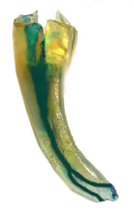
Root Curvature - Curvature of the root was found in the apical third of the root in 100 teeth (64.10%) and in the middle third in 36 teeth (23.07%). 15 teeth were found to have straight root i.e. no curvature (9.61%). In all the five teeth with two roots, both the roots were found to have curvature in the apical third. (Table 2) Amongst one rooted teeth 10 to 100 of root curvature was found in 2 teeth (1.28%), 110 and 200 in 47 teeth (30.12%), 210 to 300 in 54 teeth (34.61%), 310 or more in 33 teeth. Amongst the two rooted teeth 110 to 200 curvature was found in 4 teeth (80%) and 210 to 300 in 1 tooth (20%).
1 : Curved Root Canal
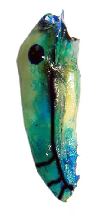
Accessory Canals – 59 teeth were found to have accessory canal (37.82%). Out of these one accessory canal was found in 41 teeth (69.49%), two accessory canals were found in 15 teeth (25.42%) and 3 or more accessory canals in 3 teeth (5.08%). Accessory canals were located in the apical third in 50 teeth (84.74%) and is the middle third in 9 teeth (15.28%)
2 : Accessory Canal
Transverse Anastamosis – 24 teeth were found to have transverse anastamosis between the canals (15.89%). In 19 teeth (79.16%) it was located in the apical third and in 5 teeth (20.83%) in the middle third
Apical Delta – Apical delta was found in 7 teeth (4.48%)
Position of Apical Foramen – Apical foramen was located at the apex or within 1 mm of apex in 129 teeth (82.69%), 1 to 2 mm distant from apex in 22 teeth (14.10%) and more than 2 mm distant from the apex in 5 teeth (5.20%).
DISCUSSION
In this study morphology of the apical portion of the root of mandibular first premolars was analyzed, since variations in the morphology play a vital role in the successful outcome of the endodontic treatment and these teeth historically have a high failure rate. (4,5,6)
Decalcification and clearing technique was chosen since it has been found to be highly effective in disclosing finer anatomic details, (8,9) though radiographs, (10) macroscopic sections,(11) 3D reconstruction,(12) computed tomography(13,14) and direct observation with microscope, (15) have been used in various studies, decalcification and clearing technique has been found to be simple, inexpensive and effective.
In this study we found two roots in 3.20% teeth which is consistent with the finding of various text (16, 17, 18, 19, 20). Invagination of the root was found in 15.74% teeth which was similar to that found by Robinson et (21) and Velumurgan and Sandhya. The root curvature was found to vary between 0 to 480 with maximum teeth (34.61%) having a curvature between 210 to 310. Among one rooted teeth, most roots had curvature in the apical third (64.10%) and 23.07% in the middle third. Among the two rooted teeth, all the roots had curvature in apical third with 80% of these having a curvature between 110to 200. None of the roots had any curvature in the cervical third.
Accessory canals were found in 37.82% teeth with their number varying from 1 to 4. Most teeth with accessory canal were found to have only one (69.49%). Most common location of these canals was the apical third (15.28% ) and none were found in the cervical third. Vertucci had reported their presence in 44.3% mandibular first premolar, which were located in the apical, middle and cervical third in 78.9%, 16.1%, & 4.3% teeth respectively.
Transverse anastamosis, intercanal connection or isthmus(22) were found in 15.89% of mandibular first premolar. These were predominantly located in the apical third (79.16% teeth) while the rest (20.83% teeth) were found in middle third. Transverse anastamosis were not found in cervical third of any tooth. Vertucci had reported their presence in 32.1% mandibular first premolar, which were located in the apical, middle and cervical third in 26.5%, 52.9% & 20.6% teeth respectively. Weller et al found intercanal connection in 30% mandibular premolar. (23)
Apical delta were found in 4.48% of the Mandibular first premolar teeth while Vertucci’s had reported their presence in 5.7%.
In this study Apical foramen were found to be located at or within 1 mm of the apex in 82.69% teeth, Velumurgan and Sandhya reported their presence in 83% teeth whereas Vertucci reported only 15% of Mandibular first premolar having apical foramen at the apex. They were found to be located 1 to 2 mm from the apex in 14.10
% teeth and 2mm or more in 3.20% teeth, Velumurgan & Sandhya found them to be 0.5 mm distant in 6%, 1 mm from apex in 9.7% & 2 mm from apex in 1.2% teeth, Vertucci had found 85% teeth with apical foramen lateral to apex.
CONCLUSION:-
It can be concluded that mandibular first premolars commonly have one root with 210 to 300 curvature and one accessory canal in the apical third, with apical foramen located within 0.5 mm of the apex. In presence of a second canal transverse anastamosis or apical delta can be expected, with apical foramen located 0.5 to 2mm distant from the apex.
REFERENCES
- Cutright DE, Bhasker SN. Pulpal vasculature as demonstrated by a new method. Oral Surg Oral Med Oral Pathol Oral Radiol Endod .1974;38:308-314.
- Mjör IA, Nordahl I. The density and branching of dentinal tubules in human teeth. Arch Oral Biol.1996;41:401-412.
- Majör IA, Simth MR, Ferrari M, Mamnoci F.The structure of dentin in the apical region of human teeth. Int Endod J.2001;34:346-353.
- Cohen S, Hargreaves KM. Pathways of the Pulp. 9ed. St. Louis.Mosby-Elsevier.2006.216
- Slowey RR. Root canal anatomy. Road map to successful endodontics. Den Clin North Am. 1979;23:555-573.
- Ingle J, Bakland L. Endodontics, 5th ed. Hamilton: BC Decker: 2008.202.
- Robertson D, Leeb J, Mckee M, Brewer E. A clearing technique for the study of root canal systems. J O E. 1980;6:421-4.
- Velumurgan & Sandhya R. Root Canal morphology of mandibular first premolar in an Indian Population: a laboratory study.Int Endod J.2009;42:54-58.
- Rahimi S, Shahi S, Yavari H, Manafi H, Eskandarzadeh N. Root canal configuration of Mandibular first & second premolars in as Iranian population. JODDD.2007; Vol 1, No. 2, 59-64.
- Pineda F, Kutter Y. Mesiodistal and Buccolingual roentogenographic investigation of 7,275 root canal .Oral Surg Oral Med Oral Pathol. 1972; 33.101-10.
- Baisden MK, Kulilid JC, Weller RN: Root canal configuration of the mandibular first premolar. J Endod. 1992; 18:505-8.
- Mikrogeorgis G, Lyrondia KL, Nikopoulos N., Pitas I, Molyvdas I, Lambrianidis TH. 3D Computer-aided reconstruction of six teeth with morphological abnormalities. Int Endo J.1999;32, 88-93.
- Caliskan MK, Pehlivan Y, Sepetcioglu F, Turkun M, Tuncer SS. Root Canal Morphology of human permanent teeth in Turkish population. JOE.1995; 21:200-4.
- Rwenyonyi CM, Kutesa AM, Muwazi LM, Buwembo W. Root and canal morphology of Maxillary first and second permanent molar teeth in a Ugandan population, Int Endod J. 2007; 40:679-83.
- Sempire HN, Hartwell GR. Frequency of second mesiobuccal canals in maxillary molars as determined by use of an operating microscope: clinical study. J O E.2000; 26:673-4.
- Ash M, Nelson S. Wheeler’s Dental Anatomy, Physiology and Occlusion. 8th Ed Philadelphia: Saunders: 2003
- Brown P, Herbranson E. Dental Anatomy & 3D Tooth Atlas version 3.0.2nd ed. Illinois. Quintessence. 2005.
- Black G. Descriptive Anatomy of teeth.4th ed. Philadelphia. SS White Dental Manufacturing Company. 1902.
- Fuller J Denehy G. Concise Dental Anatomy & Morphology, 2nd ed. Chicago Year Book Medical Publishers, Inc: 1984.
- Jordon R, Abrams L, Krans B. Krans Dental Anatomy & Occlusion. 2nd ed. St. Louis Mosby Year Book. MC. 1992
- Robinson S, Czerny C, Gahleitner A. Bernhart T, Kainberger FM. Dental CT evaluation of mandibular first premolar root configuration and canal variations. Oral Surg Oral Med Oral Pathol Oral Radiol Endod. 2000; 93:328-32.
Copyright Priory Lodge Education Limited 2010
First Published October 2010
Click
on these links to visit our Journals:
Psychiatry
On-Line
Dentistry On-Line | Vet
On-Line | Chest Medicine
On-Line
GP
On-Line | Pharmacy
On-Line | Anaesthesia
On-Line | Medicine
On-Line
Family Medical
Practice On-Line
Home • Journals • Search • Rules for Authors • Submit a Paper • Sponsor us
All pages in this site copyright ©Priory Lodge Education Ltd 1994-


