Plastic Bronchitis leading to Respiratory Failure Following the Fontan Procedure:
A pediatric case report, proposed pathophysiology and review of current treatment modalities.
(Nishant Shah MD a, Mary Baldauf MD a, Mayank Shukla MD a, Patrick Flynn MD b, Sarita Dhuper MD a )
-
a Department of Pediatrics, Brookdale University Hospital & Medical Center, 1 Brookdale Plaza, Brooklyn, NY 11212
- b Department of Pediatrics, New York Presbyterian Hospital, Weill Cornell Medical College, 525 East, 68th Street, New York, NY 10021
Abstract:
Plastic bronchitis, a rare but well known disorder was first described in 1902 . It is associated with many pulmonary and nonpulmonary conditions in which bronchial casts which resemble bronchial anatomy are produced.
We present a 4 ½ year old girl with congenital heart disease status post Fontan repair who was admitted repeatedly with respiratory distress and was diagnosed preterminally with plastic bronchitis. Here we review the likely pathophysiology as well as standard and experimental treatment modalities on the basis of an extensive literature review.
Case Report:
The patient was born through a full-term normal spontaneous vaginal delivery with no known complications. The diagnosis of a Double Inlet Single Ventricle with L-TGA was made at four months of age when she presented to the inpatient service with mild respiratory distress.
She was referred for her first cardiac catheterization at 5 months of age which revealed increased pulmonary blood flow with a Qp: Qs ratio of 4.2:1 and a mean pulmonary artery pressure of 50 mmHg with a pulmonary capillary wedge pressure of 18 mmHg. Pulmonary vascular resistance was 3 Wood units. The patient underwent a pulmonary artery banding procedure at 6 months of age. She subsequently underwent a bi-directional cavo-pulmonary (Glenn) shunt procedure at 14 months of age. At 3 ½ years of age, a lateral tunnel fenestrated Fontan was performed. Post-operatively, she developed a persistent right sided pleural effusion. Catheterization 6 months post-operatively revealed a normal pressure in the Fontan circuit, cardiac index of 3.6 litres/minute/m2 and no significant obstruction in the branch pulmonary arteries. The gradient across the bulboventricular foramen in the subaortic area was under 20 mmHg. The pulmonary vascular resistance was 2.5 Wood units. Angiography demonstrated the Fontan fenestration was patent and unobstructed. Angiography in the ascending, thoracic and abdominal aorta showed multiple small collaterals in the right lung including the intercostal arteries, which were occluded by Coil embolizations. Over time, with aggressive diuretic therapy and chest physiotherapy, her pleural effusions improved. On discharge, her EKG demonstrated sinus rhythm and the echocardiogram revealed normal ventricular function and a patent fenestration with right to left flow and no pericardial effusion. However, within a month, she again developed increasing respiratory distress, a decreased oxygen saturation levels and radiographic evidence of a large right-sided pleural effusion with areas of atelectesis and/or scarring.
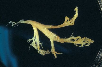
Over the next 6 months, she continued to have recurrent right-sided pleural effusions associated with respiratory distress requiring chest tube insertions. She also developed a chronic cough that was associated with expectoration of pearly white stringy materials that were later confirmed by microscopy to be bronchial casts (Figure 1). The casts were composed of mainly fibrin, mucus and scattered lymphocytes and macrophages and a culture from a representative cast was negative. A diagnosis of plastic bronchitis was made in association with the Fontan physiology. She was treated with antibiotics, steroids, albuterol and N-acetylcysteine nebulizations, oxygen and vigorous chest physiotherapy. The patient was scheduled to undergo an elective bronchoscopy to remove large bronchial casts and thoracoscopy to lyse any long-standing adhesions and to re-expand the collapsed portions of her lung. However, before the patient underwent elective bronchoscopy, she developed acute respiratory failure requiring intubation and ventilatory support. During this critical period, multiple bronchoscopies were performed to remove thick mucus plugs and bronchial casts. Unfortunately, her clinical condition continued to deteriorate, requiring high pressure ventilation and multiple inotropes. Finally, this led to her ultimate demise at the age of 4 ½ years, approximately 14 months after her Fontan procedure.
Discussion:
Plastic bronchitis has been diagnosed in a variety of pulmonary pathologies including asthma , , respiratory infections , cystic fibrosis , bronchiectasis 2 and acute chest syndrome associated with sickle cell disease . Interestingly, several non-pulmonary pathologies have also been associated with the diagnosis of plastic bronchitis. These include complex congenital cyanotic heart disease (especially after Fontan procedure), constrictive pericarditis and lymphangiomatosis .
The pathogenesis of plastic bronchitis is not well understood. There are likely two mechanisms for the development of plastic bronchitis:
(1) Injury to bronchi and/or impaired bronchial epithelial function secondary to inflammation or infection
(2) Impaired pulmonary lymphatic drainage.
Injury to bronchi and/or impaired bronchial epithelial function may be secondary to inflammation such as in asthma, or infection such as in pneumonia. Impaired epithelial function as seen in cystic fibrosis and bronchiectasis also contributes to the formation and propagation of thick, fibrinous bronchial casts. Infection and inflammation increase the content of fibrin and cellular components in the casts. In addition, bronchial epithelium becomes leaky for interstitial fluid and impairment in the clearing of bronchial secretions occurs.
Impaired lymphatic drainage of the lung may occur secondary to congenital or acquired conditions. Lymphangiomatosis, general lymphodysplasia in Turner syndromes 9, Fontan physiology and constrictive pericarditis have all been associated with impaired pulmonary lymphatic drainage. Lymphatic abnormalities associated with various pediatric cardiac conditions were very well documented by Languepin et al . In that report, abnormal lymphatic circulation was found in all three cases of plastic bronchitis out of that two were associated with cardiopathies. Hug et al demonstrated massively dilated pulmonary lymph vessels, ruptures of lymphatic vessels and the influx of chyle into the alveoli in a patient with Fontan physiology and plastic bronchitis at autopsy. It is well known that the Fontan operation can lead to systemic venous pressure hypertension as well as hepatic congestion, which in turn significantly increases the volume of lymph in the thoracic duct. A retrograde flow of chyle into the lungs and bronchi can occur only when widely dilated lymph channels in the lungs communicate freely with the thoracic duct or with transdiaphragmatic channels . Lymphatic abnormalities and endobronchial chylous reflux in various cardiopathies occurs mainly secondary to surgical trauma to the mediastinal lymph channels, injury causing pleural adhesions and systemic venous hypertension. Plastic bronchitis likely occurs as a result of isolated or combined effects of the above pathophysiologies.
Quite often, both bronchial epithelial dysfunction and impaired lymphatic drainage act in concert to lead to bronchial cast formation and plastic bronchitis.
Seear et al 3 proposed that bronchial casts can be divided into two distinct groups:
-
Inflammatory casts
-
Acellular casts
Inflammatory casts are composed primarily of fibrin with little mucin and have cellular infiltrates, particularly comprised of eosinophils. Inflammatory casts are typically seen in the plastic bronchitis of patients with predominant bronchial disease such as asthma and cystic fibrosis.
Acellular casts are composed mainly of mucin with little fibrin and no inflammatory cells except occasional mononuclear cells. This was histologically similar to our case. This type of cast is predominantly seen in patients with lymphatic drainage impairment as the major cause of plastic bronchitis, as in Fontan physiology. Some patients with plastic bronchitis may have pathological features of both groups. P. Madsen et al present a thorough review of histological classifications of plastic bronchitis.
The treatment of plastic bronchitis is focused on the mechanical removal of the thick bronchial casts and prevention of further cast formation. After an extensive review of the literature, we propose the following treatment modalities:
(1) Removal of bronchial casts and maintenance of adequate ventilation:
The standard of care includes oxygen, chest physiotherapy, bronchodilators, N-acetylcysteine 3 and bronchoscopy. Repeated bronchoscopic aspirations and removal of casts are required in acute cases. Lobectomy has resulted in cessation of cast formation particularly when a specific lobe of the lung is involved in chronic inflammation and subsequent cast production and obstruction. The uses of t-PA , Recombinant human DNase (rhDNase) and Urokinase have been documented in the treatment of plastic bronchitis. Wakeham et al reported successful long term treatment of plastic bronchitis with t-PA in Fontan patient. The patient was given t-PA every six hourly on long term basis to prevent recurrence of bronchial cast formation.
(2) Treatment and prevention of bronchial inflammation and injury:
Steroids and antibiotics have been used widely to treat plastic bronchitis with certain underlying pathologies. A therapeutic trial of low-dose and long-term oral Azithromycin showed marked clinical and radiological improvement in a healthy patient with plastic bronchitis probably secondary to its anti-inflammatory action.
(3) Prevention of endobronchial chyle reflux:High protein/low fat diets with medium-chain triglycerides have been used to reduce lymph flux and chylous effusions with variable success .
Surgical ligature of the thoracic duct is considered by some the most effective treatment for chylothorax . Languepin et al demonstrated cessation of bronchial cast formation in one pediatric patient with Fontan physiology after thoracic duct ligation.
Prevention of chyle reflux by improvement of hemodynamics in Fontan patients has been attempted through various interventions. In one case, a permanent pacemaker was placed which resulted in improvement in cardiac function with atrioventricular synchrony and resolution of cast formation.10 Stiller and colleagues described two children with plastic bronchitis following a Fontan procedure. One child was given diuretics to optimize heart function (reducing filling pressures) and the other was given subcutaneous standard heparin. These therapies led to cessation of cast formation in both patients. Percutaneous creation of a stent fenestration in the Fontan circuit resulted in full symptomatic recovery from plastic bronchitis in a patient who had unobstructed Fontan pathways and raised central venous pressure (15 mm Hg). A pericardiectomy showed immediate improvement with cessation of cast formation in a patient with Fontan physiology and secondarily pleural as well as pericardial effusions.7 The endothelin receptor antagonist, Bosentan showed clinical, exercise and hemodynamic improvement in one patient with plastic bronchitis after Fontan.
All reported modalities used to treat plastic bronchitis are summarized in Table 1. None of the above therapies, however, has proven its efficacy under evidence-based scrutiny, and the prognosis of this condition remains poor. In pediatric patients with plastic bronchitis secondary to Fontan physiology, 45% of the 18 reported cases have died from asphyxia secondary to airway obstruction (Table-2). This current patient died from airway obstruction leading to Systemic Inflammatory Response Syndrome and multiple organ system dysfunctions. Plastic bronchitis in patients with Fontan physiology is life threatening and Pediatricic Intensivists, Pulmonologists and Pediatric Cardiologists should maintain a high index of suspicion in such patients who repeatedly demonstrate respiratory pathology. A low threshold for diagnostic and well as therapeutic bronchoscopy is also warranted. Early utilization of aggressive pulmonary clearance is recommended. Considering the poor prognosis, patients with Fontan physiology who develop plastic bronchitis and fail to respond to treatment should be evaluated early for heart transplantation until a definitive therapy for plastic bronchitis is available.
Table: 1 Summary of treatment modalities used in Plastic Bronchitis:
Maintenance of adequate ventilation:
Chest Physiotherapy
Bronchodilators
N-Acetylcysteine
T-PA
Urokinase
rhDNase
Bronchoscopy
Lobectomy
Treatment and prevention of bronchial injury:
Antibiotics
Steroids
Low dose Azithromycin
Prevention of endobronchial chyle reflux:
Low fat high protein diet
Diuretics
Heparin
Bosentan, endothelin receptor antagonist
Thoracic duct ligation
Permanent pacemaker
Stent fenestration of Fontan circuit
Pericardiectomy
Table 2 Summary of Reported Cases of Plastic Bronchitis in Fontan Patients:
No |
Reference |
Age (yrs) |
Diagnosis |
Outcome |
1 |
Quasney et al ,17 |
5 |
TA |
Alive |
2 |
Seear et al, 3 |
12 |
CCCHD (BDG) |
Expired |
3 |
Seear et al, 3 |
8 |
CCCHD |
Expired |
4 |
Seear et al, 3 |
5 |
TA |
Awaiting TXS |
5 |
Bowen et al, 2 |
8 |
TA |
Alive |
6 |
3 |
TA |
Expired |
|
7 |
Costello et al, 15 |
4 |
DILV |
Expired |
8 |
6 |
CCCHD |
Expired |
|
9 |
Chaudhari et al, 24 |
3.5 |
HLHS |
Alive |
10 |
A Tzifa et al, 16 |
3.5 |
PA |
Alive |
11 |
Hug et al, 10 |
4 |
D- TGA with PA |
Expired |
12 |
5 |
CCCHD(VACTRAL) |
Alive |
|
13 |
5 |
TA |
Alive |
|
14 |
5 |
TA |
Alive |
|
15 |
3 |
PA |
Expired |
|
16 |
8 |
TAPVR, Single Ventricle |
Alive |
|
17 |
Wakeham et al 18 |
4 |
TGA, hypo. Rt. Ventricle |
Alive |
18 |
Present case |
4.5 |
DISV |
Expired |
TA- Tricuspid Atresia, BDG- Bi-directional Glenn, CCCHD- Complex Cyanotic Congenital Heart Disease, DILV- Double Inlet Left Ventricle, HLHS- Hypoplastic Left Heart Syndrome, PA- Pulmonary Atresia, D-TGA- D Transposition of Great Arteries, DISV- Double Inlet Single Ventricle, TAPVR- Total Anomalous of Pulmonary Venous Return, TS- Tricuspid stenosis
Figure: 1
REFERENCES:
- Bettmann M. Report of a case of fibrinous bronchitis, with a review of all cases in the literature. Am J Med Sci 1902; 123: 304-329
- Bowen A D, Oudjhane K, Odagiri K, Liston S L, Cumming W A, Oh K S. Plastic bronchitis: large branching mucoid bronchial casts in children. Amer J of Roentgenology. 1985 Feb; 144(2): 371-375
- Seear M, Hui H, Magee F, et al. Bronchial casts in children: A proposed classification based on nine cases and review of the literature. Am J Respi Crit Care Med 1997; 155: 364-370
- Jett JR tazelaar HD, Keim LW, Ingrassia, III TS Plastic Bronchitis: an old disease revisited. Mayo Clin Proc 1991; 66: 305-31
- Waring WW, Brunt CH, Hilman BC. Mucoid impaction of the bronchi in cystic fibrosis. Pediatrics. 1967; 39: 166-175
- Raghuram N, Pettignano R, Gal AA, et al. Plastic Bronchitis: an unusual complication associated with sickle cell disease and the acute chest syndrome. Pediatrics. 100 July 1997: 139-142
- Muller W, Von Der Hardt H, Rieger CH. Idiopathic and symptomatic plastic bronchitis in childhood. A report of three cases and review of the literature. Respiration. 1987; 52: 214-220
- Nair LG, Kurtz CP. Lymphangiomatosis presenting with bronchial cast formation. Thorax. 1996; 51: 765-766
- Languepin J, MD, Scheinmann P, MD, Mahut B, MD, Bourgeosis M L, MD, Jaubert F, MD, Brunelle F, MD, Sidi D, MD, Blic J D, MD. Bronchial casts in children with cardiopathies: The rold of pulmonary lymphatic abnormalities. Pediatr Pulmonol. 1999; 28: 329-336
- Hug M I, Ersch J, Moenkhoff M, Burger R, Fanconi S, Bauersfeld U. Circulation. 2001; 103: 1031
- Cromme-Dijkhuis AH, Hess J, Hahlen K, Henkens CM, Bink-Boelkens MT, Eygelaar AA, Bos E. Specific sequele after fontan operation at mid and long term followup. J Thorac Cardiovasc Surg 1993; 106: 1126-1132
- Maier HC. Chylous reflux in the lungs and pleura. Thorax 1968; 23: 281-296
- Madsen P, Shah Samir A, Rubin Bruce K, Plastic bronchitis: new insights and a classification scheme, Pediatric Respiratory Reviews. 2005; 6: 292-300
- Park J Y, Elshami A A, Kang D S, Jung T H. Plastic bronchitis. Eur Respir J. 1996; 9: 612-614
- Costello JM, Steinhorn D, McColley S, at al. Treatment of plastic bronchitis in a fontan patient with tissue plasminogen activator: A case report and review of the literature. Pediatrics Vol. 109 No.4 April 2002: 67
- Tzifa A, Robards M, Simpson J M. Plastic bronchitis; a serious complication of the Fontan operation International Journal of Cardiology, 2005; 101: 513-514
- Quasney MW, Orman K, Thompson J, et al. Plastic bronchitis occurring late after the fontan procedure: treatment with aerosolized urokinase. Crit Care Med. 2000; 28: 2107-2111
- Martin K. Wakeham, MD; Andrew H. Van Bergen, MD; Luis E. Torero, MD; Javeed Akhter, MD. Long term treatment of plastic bronchitis with aerosolized tissue plasminogen activator in a Fontan patient. Pediatr Crit Care Med 2005 Vol. 6,No. 1: 76-78
- Onoue Y, Adachi Y, Ichida F, Miyawaki T. Effective use of corticosteroid in a child with life-threatening plastic bronchitis after Fontan operation. Pediatr Intemational 2003; 45: 107-109
- Schultz K D, Oermann C M. Treatment of cast bronchitis with low dose oral azithromycin. Pediatr Pulmonol. 2003; 35: 139-143
- Bond SJ, Guzzetta PC, Snyder ML, Randolph JG. Management of pediatric postoperative chylothorax. Ann Thorac Surg 1993; 56: 469-473
- Marts BC, Naunheim KS, Fiore AC, Pennington DG. Conservative versus surgical management of chylothorax. Am J Surg 1992; 164: 532-535
- Stiller B, Riedel F, Paul K, Van Landeghem FKH (2002) Plastic bronchitis in children with Fontan palliation: analogue to protein losing enteropathy. Pediatr Cardiol 23: 90-94
- Chaudhari M, Stumper O. Plastic bronchitis after Fontan operation: treatment with stent fenestration of the Fontan circuit. Heart 2004; 90: 801
- Sotiria C. Apostolopoulou, MD, PhD, John Papagiannis, MD, and Spyridon Rammos, MD, PhD Bosentan induces clinical, exercise and hemodynamic improvement in a pre-transplant patient with plastic bronchitis after Fontan operation. J Heart Lung Transplant 2005; 24: 1174-1176
- Duncan W, Tyrrell M, Bharadwaj B, George D. Fontan’s operation complications. Pediatr Cardiol. 1993; 14: 62-63
- Peleg U, Schwartz S, Sirota G, Hochman I, Cohen D, Picard E. Persistent plastic bronchitis in a child after cardiac surgery. IMAJ 2005; 7: 122-124
- Wilson J, Russell J, Williams W, Benson L. Fenestration of the Fontan circuit as treatment for plastic bronchitis Pediatric Cardiology; August, 2005.
- Brogan TV, Finn LS, Pyskaty DJ et al. Plastic Bronchitis in children: a case series and review of the medical literature. Pediatr Pulmonol 2002; 34: 482-487
- Barber BJ, Burch GH, Tripple D, Balaji S. Resolution of plastic bronchitis with atrial pacing in a patient with Fontan physiology. Pediatr Cardiol 2004; 25: 73-76
- Mcmahon CJ, Nihill MR, Reber A. The bronchial cast syndrome after the Fontan procedure: further evidence of its etiology. Cardio Young 2001; 11: 345-351
- Onoue Y, Adachi Y, Ichida F, Miyawaki T. Effective use of corticosteroid in a child with life-threatening plastic bronchitis after Fontan operation. Pediatr Intemational 2003; 45: 107-109
Expect Additional Medical Journals to appear soon
- Evidence Based Medicine On-Line (tm)
- Medical Ethics On-Line (tm)
- Paediatrics (Pediatrics) On-Line (tm)
- Obstetrics On-Line (tm)
- Gynaecology On-Line (tm)
- Microbiology On-Line (tm)
- Cardiology On-Line (tm)
- Surgery On-Line (tm)
- Pharmacology On-Line (tm)
- Psychology On-Line(tm)
- Health Management On-Line (tm)
- Nurse On-Line (tm)
Directors: Ben Green, Rob Glenning.
A UK Limited Company
All pages copyright ©Priory Lodge Education Ltd 1994-